Hip & Knee
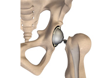
Hip Anatomy
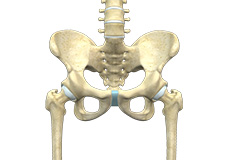
The hip joint is the largest weight-bearing joint in the human body. It is also referred to as a ball and socket joint and is surrounded by muscles, ligaments and tendons. The thighbone or femur and the pelvis join to form the hip joint.
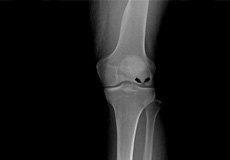
Knee Anatomy
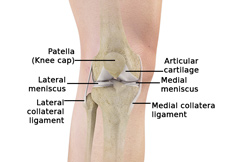
The knee is a complex joint made up of different structures including bones, tendons, ligaments and muscles. They all work together to maintain normal function and provide stability to the knee during movement.
Osteoarthritis
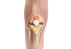
Osteoarthritis, also called degenerative joint disease, is the most common form of arthritis. It occurs most often in older people. This disease affects the tissue covering the ends of bones in a joint (cartilage). In a person with osteoarthritis, the cartilage becomes damaged and worn out causing pain, swelling, stiffness and restricted movement in the affected joint.
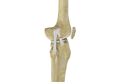
ACL Tears
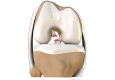
The anterior cruciate ligament, or ACL, is one of the major ligaments of the knee that is in the middle of the knee and runs from the femur (thighbone) to the tibia (shinbone). It prevents the tibia from sliding out in front of the femur. Together with posterior cruciate ligament (PCL) it provides rotational stability to the knee.
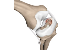
Meniscal Tears
Meniscus tear is the commonest knee injury in athletes, especially those involved in contact sports. A sudden bend or twist in your knee causes the meniscus to tear. This is a traumatic meniscus tear. Elderly people are more prone to degenerative meniscal tears as the cartilage wears out and weakens with age. The two wedge-shaped cartilage pieces present between the thighbone and the shinbone are called meniscus.
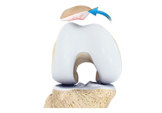
Patellar Dislocation/Patellofemoral Dislocation
Patella (kneecap) is a protective bone attached to the quadriceps muscles of the thigh by quadriceps tendon. Patella attaches with the femur bone and forms a patellofemoral joint. Patella is protected by a ligament which secures the kneecap from gliding out and is called as medial patellofemoral ligament (MPFL).
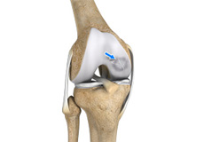
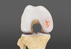
Osteochondritis Dissecans
The knee, mostly the femoral condyles are most commonly affected. The two femoral condyles make up for the rounded end of femur (thighbone). Each knee has two femoral condyles, the medial femoral condyle on the inside of the knee and the lateral femoral condyle on the outside of the knee. Osteochondritis dissecans occurs within the lateral aspect of the medial femoral condyle.
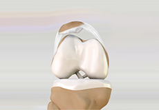
Lateral Patellar Compression Syndrome
Lateral patellar compression syndrome refers to pain under and around your kneecap. It is a common complaint among runners, jumpers, and other athletes such as skiers, cyclists and soccer players.
The patella, also called kneecap, is a small flat triangular bone located at the front of the knee joint.
MCL Sprain
The medial collateral ligament (MCL), a band of tissue present on the inside of your knee joint, connects your thighbone and shinbone (bone of your lower leg). The MCL maintains the integrity of the knee joint and prevents it from bending inward.
Your MCL may get sprained or injured while twisting, bending or quickly changing direction.
Other Conditions
Articular Cartilage Defects
Articular or hyaline cartilage is the tissue lining the surface of the two bones in the knee joint. Cartilage helps the bones move smoothly against each other and can withstand the weight of the body during activities such as running and jumping. Articular cartilage does not have a direct blood supply to it so has less capacity to repair itself. Once the cartilage is torn it will not heal easily and can lead to degeneration of the articular surface, leading to development of osteoarthritis.
Patellofemoral Instability
The knee can be divided into three compartments: patellofemoral, medial and lateral compartment. The patellofemoral compartment is the compartment in the front of the knee between the kneecap and thighbone. The medial compartment is the area on the inside portion of the knee, and the lateral compartment is the area on the outside portion of the knee joint. Patellofemoral instability means that the patella (kneecap) moves out of its normal pattern of alignment.
Knee Angular Deformities
Angular deformities of the knee are common during childhood and usually are variations in the normal growth pattern. Angular deformity of the knee is a part of normal growth and development during early childhood.
Osteonecrosis of the Knee
Osteonecrosis is a condition in which death of a section of bone occurs because of lack of blood supply to it. It is one of the most common causes of knee pain in older women. Women over the age of 60 years of age are commonly affected, three times more often than men.
Non-surgical Treatments
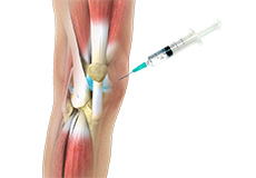
Viscosupplementation
Viscosupplementation refers to the injection of a hyaluronan preparation into the joint. Hyaluronan is a natural substance present in the joint fluid that assists in lubrication. It allows smooth movement of the cartilage covered articulating surfaces of the joint.
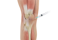
Cortisone Injection
Cortisone is a corticosteroid released by the adrenal gland in response to stress and is a potent anti-inflammatory agent.
Cortisone injections are recommended in injuries that cause pain and inflammation, and those that don’t require surgical treatment. One such condition is frozen shoulder.
Surgical Treatments
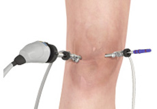
Knee Arthroscopy
The knee joint is one of the most complex joints of the body. The lower end of the thighbone (femur) meets the upper end of the shinbone (tibia) at the knee joint. A small bone called the patella (kneecap) rests on a groove on the front side of the femoral end. A bone of the lower leg (fibula) forms a joint with the shinbone.
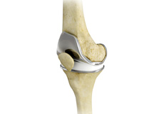
Total Knee Replacement
The knee is made up of the femur (thighbone), the tibia (shinbone), and patella (kneecap). The meniscus, the soft cartilage between the femur and tibia, serves as a cushion and helps absorb shock during motion. Arthritis (inflammation of the joints), injury, or other diseases of the joint can damage this protective layer of cartilage, causing extreme pain and difficulty in performing daily activities. Osteoarthritis is a type of arthritis.
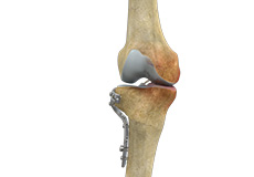
Knee Osteotomy
Knee osteotomy is a surgical procedure in which the upper shinbone (tibia) or lower thighbone (femur) is cut and realigned. It is usually performed in arthritic conditions affecting only one side of your knee and the aim is to take pressure off the damaged area and shift it to the other side of your knee with healthy cartilage. During the surgery, your surgeon will remove or add a wedge of bone either below or above the knee joint depending on the site of arthritic damage.
Knee Reconstruction
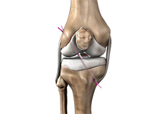
The knee is the most complex joint in the body and is formed by the articulation between the thighbone (femur) and the shinbone (tibia). A kneecap is present over the front of the joint to provide extra protection. These bones are held together by four strong rope like structures called ligaments. Two collateral ligaments are present on either side of the knee and control the sideway movements of the knee. The other two ligaments are the anterior and posterior cruciate ligaments, ACL and PCL respectively, which are present in the center of the knee joint and cross each other to form an “X”.
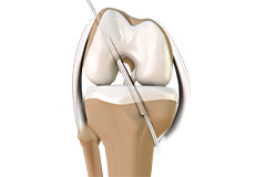
ACL Reconstruction (Patellar and Hamstring Tendon)
The anterior cruciate ligament is one of the major stabilising ligaments in the knee. It is a strong rope like structure located in the centre of the knee running from the femur to the tibia. When this ligament tears unfortunately, it does not heal and often leads to the feeling of instability in the knee.
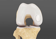
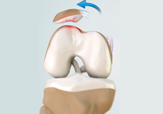
Cartilage Restoration
Articular Cartilage is the white tissue lining the end of bones where these bones connect to form joints. Cartilage acts as cushioning material and helps in smooth gliding of bones during movement. An injury to the joint may damage this cartilage which cannot repair on its own. Cartilage can be damaged with increasing age, normal wear and tear, or trauma. Damaged cartilage cannot cushion the joints during movement and the joints may rub over each other causing severe pain and inflammation.
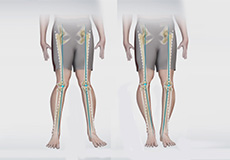
Knee Angular Deformities Correction Surgery
Angular deformities of the knee are common during childhood and usually are variations in the normal growth pattern. Angular deformity of the knee is a part of normal growth and development during early childhood. Physiologic angular deformities vary with age as:
- During first year: Lateral bowing of tibia
Total Hip Replacement
Arthritis is inflammation of the joints resulting in pain, swelling, stiffness and limited movement. Hip arthritis is a common cause of chronic hip pain and disability.
Common Types of Arthritis
- Osteoarthritis: It is characterised by progressive wearing away of the cartilage of the joint.